Bilateral Total Knee Replacement for Advanced Osteoarthritis: A Patient Case Study

Bilateral Total Knee Replacement for Advanced Osteoarthritis
Knee osteoarthritis is a degenerative joint condition in which the protective cartilage inside the knee gradually wears away. When osteoarthritis affects both knees, patients may experience persistent pain, stiffness, reduced stability and difficulty performing everyday activities.
If medication, physiotherapy, lifestyle modifications and other non-surgical treatments no longer provide adequate relief, bilateral total knee replacement may be considered.
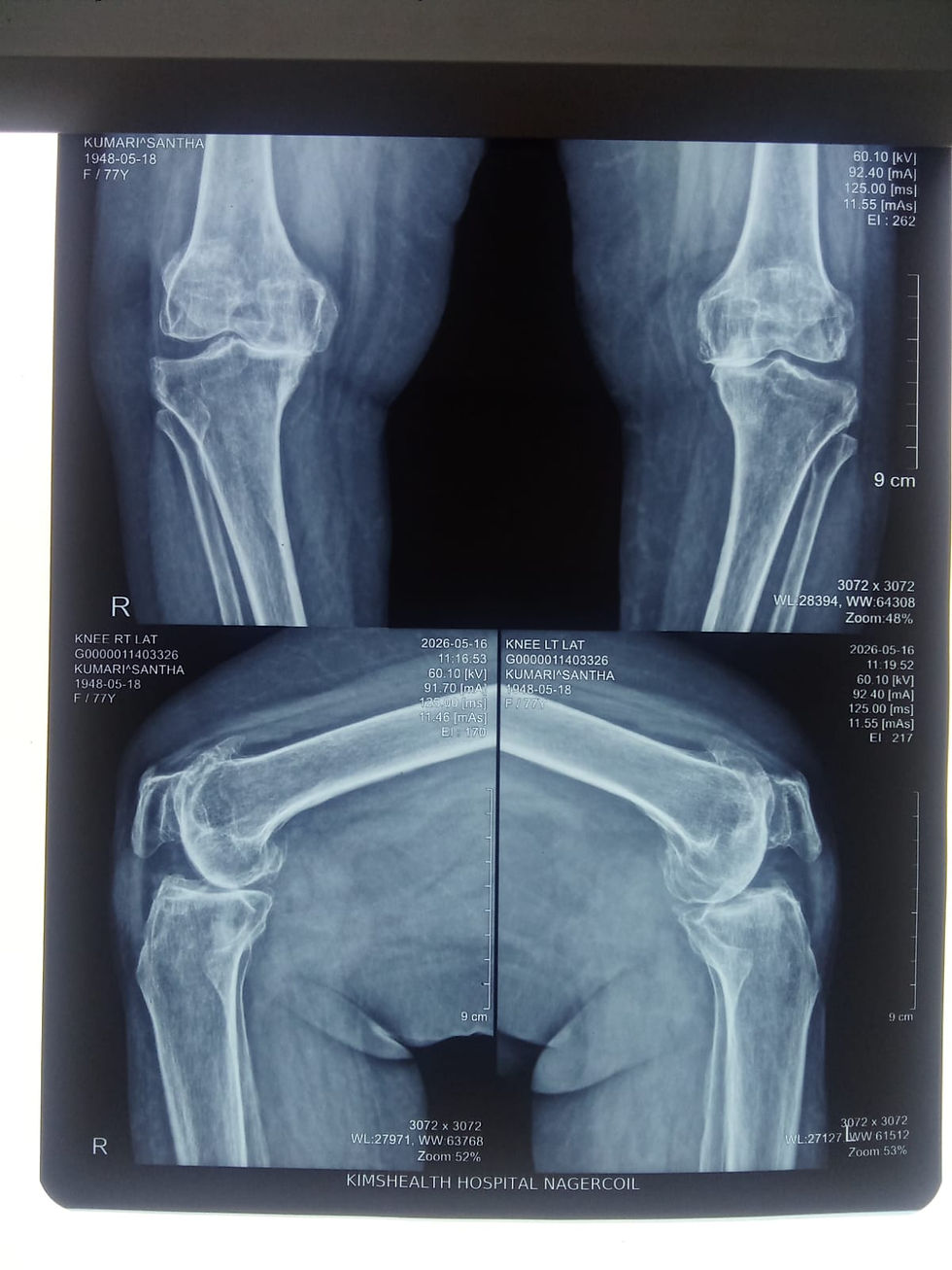
Patient’s Condition
A 74-year-old woman presented with advanced degenerative changes affecting both knee joints. The condition caused knee pain, stiffness and reduced mobility, making routine activities increasingly difficult.
Clinical examination and X-rays revealed significant deterioration of the joint surfaces in both knees. Based on the severity of the osteoarthritis and its impact on her daily life, bilateral total knee replacement was recommended.
Treatment Decision
The main objectives of the treatment were to:
Remove the damaged joint surfaces
Correct the alignment of both knees
Reduce pain and stiffness
Improve joint stability
Restore comfortable knee movement
Help the patient regain mobility and independence
Bilateral Total Knee Replacement Procedure
Bilateral total knee replacement, also known as bilateral total knee arthroplasty, was performed to treat the damaged joint surfaces in both knees.
During the procedure, the worn portions of the femur and tibia were replaced with prosthetic components designed to restore knee alignment, stability and smoother joint movement.
The postoperative X-rays show the femoral and tibial components in satisfactory position and alignment in both knees.

Postoperative Knee X-rays
The X-rays document the condition of both knee joints and the alignment of the knee replacement components following surgery. Front and side views allow the surgeon to assess implant positioning and overall joint alignment.
Postoperative X-rays form part of the patient’s follow-up evaluation and should always be interpreted together with clinical examination and recovery progress.


Comments